


Toxoplasma gondii Detection in Fecal Samples from Domestic Cats (Felis catus) in Hawai‘i1
The presence of large numbers of free-ranging feral cats (Felis catus) has raised concern in terms of both native species predation and potential disease transmission in Hawai‘i. A disease of particular concern is toxoplasmosis, caused by Toxoplasma gondii, a zoonotic protozoan parasite. We tested soil samples and cat fecal samples from cat colonies from an urban university campus and a natural, coastal ecosystem for T. gondii oocysts using standard molecular procedures. Soil and fecal samples were collected from cat colony sites at the University of Hawai‘i at Mānoa (UHM), and additional fecal samples were collected from cats trapped within Ka‘ena Point Natural Area Reserve (KPNAR). Toxoplasma gondii DNA was detected in 5% (3 of 60) of fecal samples from UHM, but no T. gondii DNA was detected from soil samples. At KPNAR, 22.2% (2 of 9) of fecal samples were positive for T. gondii DNA. Presence of T. gondii at the university study sites suggests that cat colonies may be a potential health hazard for landscaping personnel, students, staff, and visitors. Likewise, presence of T. gondii at KPNAR, and potentially other coastal habitat(s) for ground-nesting seabirds or marine mammals, also suggests a disease risk and should be considered when managing those areas. Toxoplasmosis is a growing concern to both people and wildlife, and further work is needed to determine pathways of transmission both within and between terrestrial and marine ecosystems of Hawai‘i.
cat colony, Felis catus, natural area reserve, oocyst, PCR, Toxoplasma gondii, toxoplasmosis
Toxoplasma gondii is a tissue–cyst-forming protozoan parasite that can infect and cause disease (i.e., toxoplasmosis) in all warm-blooded animals (Dubey 2010). The life cycle of T. gondii consists of both sexual and asexual stages, with members of the cat family (Felidae) being the only known definitive hosts in which the sexual life stage occurs. Asexual, clonal reproduction can also occur in felids, presenting a unique opportunity for the parasite to undergo sexual reproduction followed by clonal amplification (Wendte et al. 2010). [End Page 501] When this occurs, a more virulent strain of T. gondii may be produced and shed by the felid definitive host as highly stable, environmentally resistant oocysts (Dubey and Jones 2008), thus increasing the risk of subsequent disease outbreaks in exposed intermediate hosts (Wendte et al. 2010, Wendte et al. 2011). Once oocysts are shed and sporulate in the environment, transmission occurs through ingestion of oocysts in soil, grass, fruits, and vegetables; inhalation; or in contaminated water (Teutsch et al. 1979, Tenter et al. 2000, Dubey and Jones 2008, Dubey 2010). Further, research has shown that oocysts shed by the definitive felid host can wash into coastal marine waters, resulting in disease of naive marine mammals (Shapiro et al. 2012; VanWormer, Conrad et al. 2013; Shapiro et al. 2014, VanWormer et al. 2016).
Toxoplasma gondii in domestic cats (Felis catus) is of particular concern because people interact with cats both as pets and as a nuisance species. Free-ranging cats tend to have higher instances of toxoplasma infection than pet cats, due to their diet of wild prey (Dubey and Jones 2008, Dubey 2010; VanWormer, Fritz, et al. 2013). Cats contract T. gondii either by ingesting oocysts deposited in the environment or through ingestion of tissue-cysts in prey that are intermediate hosts for this protozoan parasite. Transmission of T. gondii can also occur congenitally. Transmission of T. gondii to other warm-blooded intermediate hosts, both wild and domestic, occurs through similar mechanisms of direct ingestion of oocysts, tissue-cysts, or congenital infection (Dubey et al. 1986).
As with most oceanic islands, felids did not occur in Hawai‘i until European explorers and settlers introduced domesticated cats (Berger 1981), which are now widespread throughout the main Hawaiian Islands as both pets and free-ranging animals in both rural and urban areas (Amarasekare 1994; Lohr, Cox, and Lepczyk 2013). The high abundance of cats in Hawai‘i combined with its favorable climate for the survival of T. gondiioocysts has important implications for the archipelago’s already decreasing native wildlife abundance (Duffy and Capece 2012; Lohr, Cox, and Lepczyk 2013) because oocysts entering the environment through felid feces are likely to become part of freshwater runoff into streams and nearshore areas (Miller et al. 2008; VanWormer, Conrad, et al. 2013; VanWormer et al. 2016). Though transmission pathways in Hawai‘i are poorly understood, morbidity and mortality associated with T. gondii have been identified in a number of native Hawaiian animals, including endangered species [Hawaiian crow, Corvus hawaiiensis (Work et al. 2000); Hawaiian goose, Branta sandvicensis (Work et al. 2002, 2016); and Hawaiian monk seal, Monachus schauinslandi (Hawai‘i Department of Health 2000, Honnold et al. 2005, Barbieri et al. 2016)]; spinner dolphin, Stenella longirostris (Migaki et al. 1990); red-footed booby, Sula sula (Work et al. 2002); and in livestock (Dubey and Jones 2008). As in other island systems, the lack of native felids in Hawai‘i indicates that the native wildlife did not evolve with T. gondii and therefore may be naive, and thus more susceptible, to T. gondii–related pathology, morbidity, and mortality (Hollings et al. 2013). Instances of T. gondii infection in marine mammals in Hawaiian coastal areas suggests that T. gondii transmission may be occurring through freshwater runoff containing oocysts, which has further implications for other wildlife as well as ocean recreation users (Miller et al. 2008).
Despite the documented risk of T. gondii infection and subsequent morbidity and possible mortality in native, naive Hawaiian wildlife, there are few studies that report environmental contamination with T. gondii by nonnative felids. For example, on Mauna Kea, a volcano on Hawai‘i Island with forests harboring many endangered bird species, 37.3% of cats were seropositive for T. gondii (Danner et al. 2007). On the island of O‘ahu, 0.5% of feral cats tested were shedding oocysts and 20% had antibodies for T. gondii (Wallace 1971). Further, the transmission pathways by which native Hawaiian wildlife, both terrestrial and marine, become infected with T. gondii remain unknown. As such, further research is warranted to fully appreciate the transmission dynamics and full impact of this highly infectious, potentially deadly protozoan parasite within the fragile terrestrial and marine ecosystems of Hawai‘i. Therefore, our objective was to investigate environmental [End Page 502] contamination with T. gondii in soil and cat fecal samples from cat colonies located on a densely populated urban university campus and in cat fecal samples from an uninhabited coastal reserve.

Map of the University of Hawai‘i at Mānoa with sampling locations and inset showing KPNAR.
materials and methods
Site Description
We sampled cat colonies on the island of O‘ahu at the University of Hawai‘i at Mānoa (UHM) main campus in Honolulu, Hawai‘i, and feral cats from Ka‘ena Point Natural Area Reserve (KPNAR). Both cat feces and environmental (soil) samples were collected for molecular detection of T. gondii.
The UHM is an urban campus that supports ~14 total cat colonies, of which we randomly selected four for sampling (Figure 1). These four sites varied in both the type of vegetation and ground cover as well as the number of cats observed. Numbers at the sites ranged from 13 to 36 cats, on average, with all sites having areas of vegetation that the cats used for resting and shelter. Cat numbers [End Page 503] are based on observations between 1800 hours and 1900 hours on days with favorable weather as part of a regular monitoring program (Davis and Lepczyk 2010).
The KPNAR was used as a natural area in comparison to the urban UHM campus. This natural area reserve is approximately 0.24 km2 and is located at the northwestern end of the Wai‘anae Mountain Range on O‘ahu Island, Hawai‘i. The natural area is a breeding location for Laysan albatrosses (Phoebastria immutabilis) and wedge-tailed shearwaters (Puffinus pacificus) and is frequented by the critically endangered Hawaiian monk seal (Monachus schauinslandi). All nonnative animals were removed from the area within a predator-proof fence in 2012 as part of the establishment of a predator-free Natural Area Reserve site (Young et al. 2013). Nine cats were removed and frozen for later analysis (Lohr, Young, et al. 2013).
Sample Collection and Processing
Soil for much of the university campus, including the sample sites, is an inceptisol from the Makiki stony clay loam series with 0% to 3% slope (Natural Resource Conservation Service 2009). However, because most of the grounds have been modified through construction and landscaping, the soil at the four sites included in this study is highly mixed. A total of 30 soil samples was randomly collected from a 1 m by 1 m grid sampling scheme at each of four colonies from the main campus, yielding a grand total of 120 soil samples. We collected soil up to approximately 10 cm depth using a surface soil probe. Collected soil also included rocks (>2 mm diameter), plant material, and surface organic matter because T. gondii oocysts could potentially become part of any aggregate containing such material. Oocyst recovery was attempted from soil samples using a sucrose flotation method based on methodology described by Lélu et al. (2011). Specifically, ~20 g of soil was added to a 50 ml cone centrifuge tube along with 20 ml of deionized water and then vortexed for 2 min to homogenize and disperse water-soluble aggregates. Samples were then centrifuged at 1,500 × g for 10 min, followed by discarding of the supernatant, and the addition of 20 ml of sucrose solution with a specific gravity (SG) of 1.15 [T. gondii oocysts have a density of 1.10 SG (Dubey et al. 1970)]. The sample was then vortexed for 2 min to ensure thorough mixing and centrifuged for 10 min (1,500 × g). The supernatant was poured equally into two 50 ml cone centrifuge tubes and 35 ml of deionized water was added, bringing the density of the solution to 1.05 SG. Samples were centrifuged for 10 min at 1,500 × g to pelletize oocysts, and the supernatant from each centrifuge tube was poured equally into another 50 ml cone centrifuge tube (four cone centrifuge tubes per sample) along with 20 ml of deionized water to bring the solution density to near 1.00 SG. Samples were shaken and centrifuged for 10 min at 1,500 × g. Supernatant from each tube was carefully discarded, ensuring that the pellet remained. The pellet from each 50 ml cone centrifuge tube was pooled, and 200 μl of the pellet was reserved in a 2.0 ml microcentrifuge tube and stored at −20°C for DNA extraction.
Defecation sites at each colony on the university campus were targeted, and 15 fecal samples were collected from each colony, for a total of 60 samples. Fecal samples were both wet and dry, though attempts were made to collect as many wet samples as possible before collecting dry samples. All fecal samples were collected on the same day and double-bagged in zip-top bags. Approximately 200 mg of feces was placed into 2 ml microcentrifuge tubes for DNA extraction and stored at −20°C until use.
Cat carcasses from KPNAR were thawed in October 2012 for gut content analysis (Lohr, Young, et al. 2013) and removal of feces. Fecal samples were collected from the lower large intestine of nine cats culled as part of a multispecies eradication of introduced mammals (Young et al. 2013). We removed all feces from each cat, each of which had >200 mg, the minimum required for analysis. Fecal samples collected from KPNAR were stored at 4°C until approximately 200 mg of each sample was placed into 2 ml microcentrifuge tubes and stored at −20°C until use for DNA extraction. [End Page 504]
Molecular Analysis
DNA (from pelleted soil and feces) was extracted using the commercial kit QIAGEN QIAamp DNA Stool Mini Kit (QIAGEN, USA) according to manufacturer’s instructions (QIAGEN 2010). DNA elute of each sample was stored at −20°C. Toxoplasma gondii–specific detection was conducted by polymerase chain reaction (PCR) targeting the GRA6 gene using the primers reported by Fazaeli et al. (2000). The amplification mixture contained 3.0 μl 5x GoTAQ buffer (Promega), 1.5 μl 1% bovine serum albumin, 1.2 μl MgCl2 (25 mM), 0.3 μM of each primer, 2.4 μl dNTP (0.2 mM each dNTP), 0.2 μl (1 unit) TAQ polymerase (Promega), and 1 μl DNA template for a reaction mixture volume of 15 μl. PCR amplification was performed for 30 cycles (MJ Research DNA Engine PTC-200). Denaturation occurred at 95°C for 2 min and 93°C for 1 min, annealing occurred at 60.8°C for 30 sec, and elongation occurred at 72°C for 1 min. PCR products were examined by electrophoresis using 1.5% agarose gel stained with ethidium bromide and visualized under ultraviolet light (UV). Samples were compared using T. gondii RH strain (Advanced Biotechnologies Inc., USA) as a positive control.
Extracted DNA from fecal samples that were amplified using the GRA6 primers were further analyzed to verify that the amplicon was T. gondii DNA. Specifically, previously published pan-apicomplexan primers that amplify across the internal transcribed space (ITS-1) anchored between the 18S and 5.8S small subunit (SSU) rDNA gene arrays were used (Gibson et al. 2011) to amplify samples that were positive using the GRA6 primers. The PCR setup and amplification (i.e., thermocycler settings) were done according to Gibson et al. (2011). Toxoplasma gondii DNA was used as a positive control and molecular grade water as the negative control. PCR products were visualized using 1% agarose gel stained with GelRed Nucleic Acid Gel Stain (Biotium Inc., Fremont, California). Amplicons were cleaned using ExoSAP-IT (Affymetrix, ThermoFisher Scientific, USA) according to the manufacturer’s protocol. Sequencing (Sanger) of all positive samples that amplified at the ITS-1 locus was performed in the Genetics Laboratory of the Washington Department of Fish and Wildlife. Sequence traces were visualized, manually quality checked, and trimmed accordingly using FinchTV (Geospiza, PerkinElmer, Akron, Ohio). Sequence identity was determined via a nucleotide BLAST search in GenBank (https://blast.ncbi.nlm.nih.gov/Blast.cgi).
results
A total of 60 fecal samples and 120 environmental samples was collected from UHM, and nine fecal samples were collected from feral cats culled at KPNAR. Of these, five samples from UHM (8.3%, 95% CI 2.7–18.4) and two from KPNAR (22.2%, 95% CI 2.8–60) amplified using the GRA6 primers (Table 1). Both locations at UHM where T. gondii was isolated also had kittens present. Toxoplasma gondii DNA was not amplified from any of the environmental soil samples. Approximate amplicon size using the GRA6 primers was 650 base pairs. All seven samples that were PCR positive with GRA6 primers also amplified using the ITS-1 primers. Sequences of the ITS-1 region of five of these samples confirmed the presence of T. gondii DNA. One sample from UHM was identified as Cytauxzoon felis, a protozoal parasite common in domestic cats. One sample that amplified at both the GRA6 and ITS-1 regions did not provide a clean sequence. Table 1 provides information on amplicon size and sequence results.
discussion
Protozoal DNA was found in 8.3% (5 of 60) of fecal samples tested from cat colonies at UHM and in 22.2% (2 of 9) of fecal samples from feral cats culled from KPNAR. Of these, three from UHM and two from KPNAR were confirmed to be T. gondii. One from UHM was confirmed to be C. felis, a protozoal parasite that is only known to infect felids and does not pose a threat to humans or other wildlife species. The occurrence of T. gondii DNA in scats was thus 5% (3 of 60, 95% [End Page 505] CI 1–13.9) at UHM and 22% (2 of 9) at KPNAR. Notably these percentages may be an underestimation of the prevalence of T. gondii because the initial PCR was done using primers for GRA6; there is only one copy of GRA6 in the genome of T. gondii (Labruyere et al. 1999). Follow-up PCR was done using ITS-1 primers, of which there are 110 copies in the T. gondii genome (Guay et al. 1993). Given the relative copy number of these two markers, screening all samples with ITS-1 primers may provide more positive results.
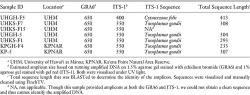
Amplicon Size from GRA6 and ITS-1 PCRs and Sequence Results of ITS-1 Amplicons from Fecal Samples Collected from Cats (Felis catus) at Two Locations in Hawai‘i
Though five samples from UHM amplified at both the GRA6 and ITS-1 regions and had amplicons of appropriate size for T. gondii, we were unable to get clean sequences for these samples across the entire amplicon (~550 bp). In general, sequence quality was poor in these samples, which is to be expected when dealing with scat samples due to inhibitors present in the sample before and after DNA extraction. Regardless, clean sequence traces for four samples from UHM and two from KPNAR were provided of approximately 150–300 base pairs and were consistent with T. gondii by BLAST search.
The higher occurrence of T. gondii DNA at KPNAR was not expected and may be a result of the small sample size and /or because we used feces harvested directly from the intestine. However, the presence of T. gondii DNA in feral cat feces indicates these cats may have been infected with T. gondii and likely were experiencing the acute active phase of the disease in which oocysts are shed into the environment. However, recent work has shown that sensitive molecular methods (i.e., quantitative PCR) may detect T. gondii DNA from tachyzoites (encysted parasites) in prey ingested by cats and DNA detection does not necessarily imply oocyst shedding by the feline host (Poulle et al. 2016). Because we were using nonnested end-point PCR, this is unlikely but nonetheless must be noted as a potential. Further work using quantitative PCR could be done to determine the number of oocysts being shed and provide better information on the specific concentration of T. gondii being shed by feral cats. Another explanation for the higher prevalence of T. gondii detected in the cats sampled from KPNAR is that cats at UHM obtain a large portion of their food from humans, and thus are eating canned or dry diets that have been conventionally prepared. In comparison, cats at KPNAR are a more rural population with little to no artificial diet provided by humans (Lohr, Young, et al. 2013).
Analyses of fecal samples from free-ranging domestic cats have found prevalences between 0.11% and 9% (Pena et al. 2006, Salant et al. 2007, Schares et al. 2008). Our finding of [End Page 506] 5% in cats in an urban setting around the university is consistent with these other studies. However, as previously mentioned, the percentage positive at KPNAR (22.2%) is higher than documented at other locations, possibly due to the small sample size. Regardless, the finding of T. gondii at a natural area site of high conservation value highlights the risk of transmission to native, endemic, and endangered wildlife in such locations, as has recently been noted in the Hawaiian Goose on Kaua‘i (Work et al. 2016).
Though T. gondii has been found in soil using molecular detection at prevalences between 5.4% and 17.8% in temperate environments (Afonso et al. 2008, Lass et al. 2009, Du et al. 2012), we did not detect T. gondii in the soil samples collected at UHM in this study. This lack of detection was in contrast to our expectations but may be due to several factors. First, we employed a regular grid around a cat colony instead of sampling directly beneath defecation sites, which may have reduced the probability of oocyst detection, because Afonso et al. (2008) demonstrated that positive T. gondii samples occurred only at cat defecation sites. Second, only 1% of cats are estimated to be shedding Toxoplasma oocysts at any point in time (Dubey 2010), thus limiting the chances of detection in soil, even if under a defecation spot. Third, feces containing oocysts may simply not have been incorporated into the soil due to the rate at which degradation and potential removal by overland flow occur. Finally, the mass of our sample used in analysis was lower than has been suggested in other studies (Dumètre and Dardé 2003). Thus, a larger sample size with larger amounts of soil that are sampled at defecation sites may increase the likelihood of detecting oocysts at our study sites in the future.
Our cat-sampling framework at UHM was not designed to test the cats themselves (i.e., direct sampling from euthanized/culled cats). Thus, no attempts were made to differentiate fecal samples from individual cats in each colony. However, samples from each site were collected on the same day and with the same collection criteria; attempts were made to collect fresh, nondesiccated samples to reduce both collecting multiple fecal samples from the same individual and also the likelihood of DNA degradation over time. Notably, both sites at UHM that were positive for T. gondii had kittens present.
Our results clearly demonstrate that T. gondii is present in cats, and possibly their prey (Poulle et al. 2016) at UHM. The occurrence of T. gondii DNA indicates that the parasite in some form (either tachyzoite or oocyst) is present in feral cats and thus the environment. As a zoonotic parasite, T. gondii in Hawai‘i has the potential to pose a health risk to the staff, faculty, students, and cat caregivers at UHM if they come into direct contact with infected feces or contaminated soil. Toxoplasma gondii also has the potential to enter the nearby aquatic and marine environments due to the proximity of the university to a stream, possibly infecting ocean recreation users, marine mammals, and /or birds (Miller et al. 2002, Conrad et al. 2005, Van-Wormer et al. 2016). Studies have indicated that oocysts entering the ocean through freshwater runoff may be ingested by filter-feeding invertebrates and fish (Arkush et al. 2003, Miller et al. 2008, Massie et al. 2010), indicating that there is a potential for such a relationship in Hawai‘i. The most prevalent filter-feeding and detritivore invertebrates in Hawai‘i include coral and various species of mollusks, crustaceans, and polychaete worms (DeFelice et al. 1998). Although these invertebrates are not usual prey items for marine birds and mammals that occur in Hawai‘i, they are prey for fish and cephalopods that marine birds and mammals prey upon. The people of Hawai‘i also consume many coastal fish and invertebrate species, including ‘opihi (Family Patellidae), a filter-feeding limpet that is often eaten raw, which could pose a risk given that numerous other filter feeders around the world have been demonstrated to be infected with T. gondii (Esmerini et al. 2010, Putignani et al. 2011, Aksoy et al. 2014, Zhang et al. 2014). Because of these potential infection pathways, research on how T. gondii oocysts move and are transmitted in tropical marine environments is greatly needed.
In the past, oocysts entering the environment at KPNAR could have had marked negative [End Page 507] effects on the wildlife that occurred there. Though speculation, both Laysan albatross and wedge-tailed shearwater adults and chicks may have ingested cat feces, aerosolized particles, or soil containing oocysts when excavating burrows or while engaged in normal activities and become infected. Hawaiian monk seals could have become infected from oocysts in freshwater runoff around the nearshore area. Oocysts may also have had the potential to accumulate in the prey of marine birds and mammals foraging in adjacent coastal waters (Conrad et al. 2005). Since the removal of all cats from within the predator-proof fence, it can be assumed that the risk of T. gondii exposure has decreased, though it is unknown what kind of risk might remain because of the lack of knowledge about the longevity of oocysts in coastal environments.
In conclusion, although we found T. gondii associated with cat colonies in both an urban and a natural area environment on O‘ahu, managing the presence of this potentially deadly protozoan parasite in the environment of Hawai‘i will prove difficult. Although removal of cats may stop T. gondii oocyst deposition into terrestrial environments, the oocysts can persist for years. Further, non-native, feral cat management on the Hawaiian Islands has become an extremely contentious issue (Lepczyk et al. 2011, Peterson et al. 2012). Given the high densities of cat populations in Hawai‘i’s terrestrial ecosystems, coupled with the large number of endangered native species, there is a critical need for improved understanding of not only the occurrence and prevalence of T. gondii in both the definitive felid host and also the native naive intermediate hosts, but also how this parasite may be transmitted across species and the subsequent impact on population health, especially of naive Hawaiian species in this fragile island ecosystem.
acknowledgments
We thank David T. Hafner Jr. for his support during the beginning stages of this project, Meghan Pawlowski and Nick Nguyen for their assistance in the field, and Lindsay Young and Eric VanderWerf from Pacific Rim Conservation and Homer Leong from USDA Wildlife Services for providing samples. Finally, we thank the two anonymous reviewers for comments on the draft version of the manuscript.
Footnotes
1. Funding for this project was provided by the University of Hawai‘i at Mānoa Office of Facilities and Grounds. Research was conducted under IBC No. 11-05-942-01-01 and IACUC No. EX-11-004 protocol approval.
